


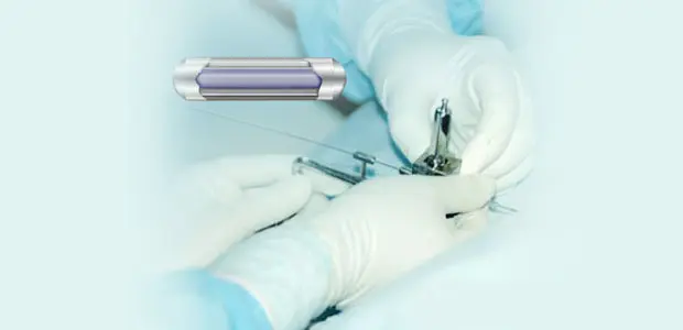
Radioactive Particle and Chemical Particle Combined Implantation
What Is Combined Particle Implantation?
Combined particle implantation is an image-guided procedure in which two different types of therapeutic “seeds” — a radioactive particle and a chemotherapy-loaded particle — are placed directly into a tumor, one after the other, using CT or ultrasound (B-mode) guidance for precise positioning. Because both particles are delivered in the same session, the tumor receives radiotherapy and chemotherapy effects at the same time, from within the tissue itself rather than from an external source.
Compared with conventional external treatment, this interstitial (inside-the-tissue) approach is generally described as minimally invasive, with a relatively short recovery and a lower rate of complications, and it is most often considered for patients whose disease is at a more advanced stage.
How the Two Particle Types Work
The Radioactive Component
The radioactive seeds most commonly used contain Iodine-125 (¹²⁵I), an isotope that emits low-energy gamma radiation capable of continuously damaging nearby tumor cells. Because the source sits inside the tumor, it can achieve a level of localized tissue destruction comparable to surgical removal, while sparing much of the surrounding healthy tissue. ¹²⁵I has a physical half-life of roughly 60 days, and its radiation output — while gradually declining — can remain biologically active inside the body for up to around six months after placement.
The Chemotherapy Component
The second particle type, sometimes called an interstitial chemotherapy implant, works differently. An anticancer drug is bound to a carrier material — either a material the body can gradually break down or one that stays intact — which is built from either naturally occurring or synthetic macromolecules. This drug-loaded carrier is placed either within the tumor mass itself or into the surrounding tissue bed, including areas left behind after surgical removal of a tumor.
Once positioned, the carrier releases the drug slowly into the immediate area, creating a local drug concentration that is markedly higher than what could ever be achieved by circulating the same drug through the bloodstream. This elevated local concentration is typically sustained for somewhere between two and fifteen days, which is intended to be long enough to damage cancer cells in that specific region and help contain the local spread of disease. Beyond its own direct effect, the chemotherapy particle is also thought to make nearby tumor cells more responsive to the radiation being delivered by the radioactive particle, effectively reinforcing the impact of both therapies at once.
How the Procedure Is Performed
The two particle types are inserted through the skin (percutaneously) using a needle, guided in real time by CT or ultrasound imaging, and are implanted either directly into the tumor or into adjacent tissue. Clinicians generally describe the procedure as straightforward to perform, with a small incision, limited tissue trauma, and a comparatively low complication rate.
Potential Advantages of Combining Both Particle Types
- Broader tissue coverage: Because the chemotherapy particle diffuses outward from its implantation point, it can expose a wider area of tissue to high drug concentrations, helping to compensate for any regions that received a lower initial radiation dose.
- Sustained local drug action: The chemotherapy particle maintains an effective concentration in the target area over an extended period, which is thought to help suppress the invasion and spread of small clusters of cancer cells; drug carried through nearby lymphatic channels may also help guard against lymph node metastasis.
- Dual mechanism of attack: By working through two distinct anticancer mechanisms simultaneously, the combination is intended to improve overall control of the tumor compared with either approach alone.
- Radiosensitizing effect: The chemotherapy component may make tumor cells more vulnerable to radiation, strengthening the effectiveness of the radioactive particle.
- More contained systemic exposure: Although some of the chemotherapy drug does eventually enter the bloodstream, it does so gradually and in small amounts. As a result, the systemic side effects are generally reported to be milder and less frequent than with an equivalent dose of standard intravenous chemotherapy.
- Single-session delivery: Because both particle types can be implanted during the same procedure, patients may be able to receive two therapeutic effects without needing a separate surgical session for each.
Which Cancers May Be Considered for This Technique
This combined implantation approach has been used as part of treatment for a range of primary tumors and metastatic disease, including cancers of the head and neck, breast, prostate, lung, liver, kidney, and cervix, as well as other solid tumors. As with any interventional oncology technique, suitability depends on tumor location, size, stage, and the patient’s overall condition, and should be assessed individually by a treating oncology team.
Determining How Many Particles Are Needed
The total number of radioactive and chemotherapy particles implanted is planned according to the size of the tumor — larger lesions generally require more particles of each type to achieve adequate coverage. Follow-up imaging is used to confirm whether particle distribution is sufficient; if coverage is found to be inadequate, additional particles may be implanted in a subsequent session.
This content is provided for general educational purposes only and does not constitute medical advice or a treatment recommendation. Outcomes vary from patient to patient, and any decision about cancer treatment should be made in consultation with a qualified oncologist based on an individual’s full diagnostic picture.