


Understanding Liver Cancer
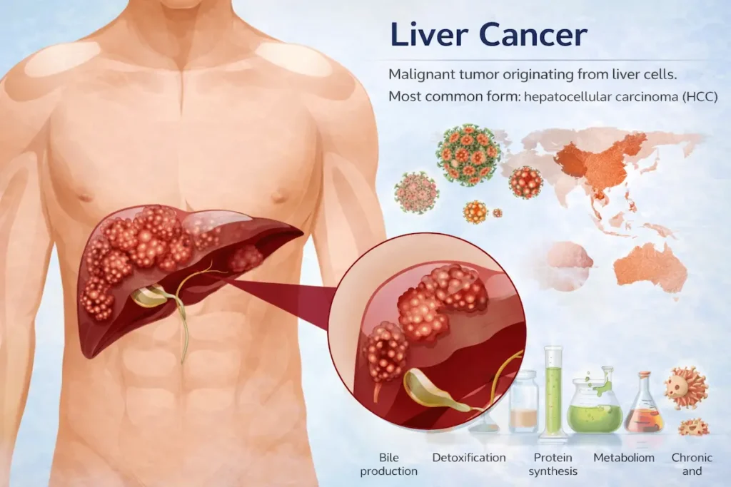
A malignant tumor originating in the hepatic tissue constitutes what is commonly referred to as liver cancer. The predominant variant is hepatocellular carcinoma (HCC), which develops from the principal functional cells known as hepatocytes. Less frequently, malignancies may emerge from the bile duct tissues or other cellular components within the liver organ.
The liver serves several critical biological functions, including metabolic processes, removal of harmful substances, production of digestive fluid, and synthesis of essential proteins. Regrettably, hepatic cancer in its initial phases typically produces minimal or absent clinical manifestations. This delayed presentation results in numerous individuals receiving their diagnosis only after the disease has progressed to advanced phases, which substantially restricts the range of therapeutic interventions available to physicians.
Geographical distribution of this malignancy reveals a concentration in regions with elevated frequencies of persistent viral inflammation of the liver, particularly throughout Southeast Asian nations and African territories. On a global scale, this condition ranks among the foremost contributors to mortality from oncological diseases.
Classification of Liver Cancer
Primary Liver Carcinoma
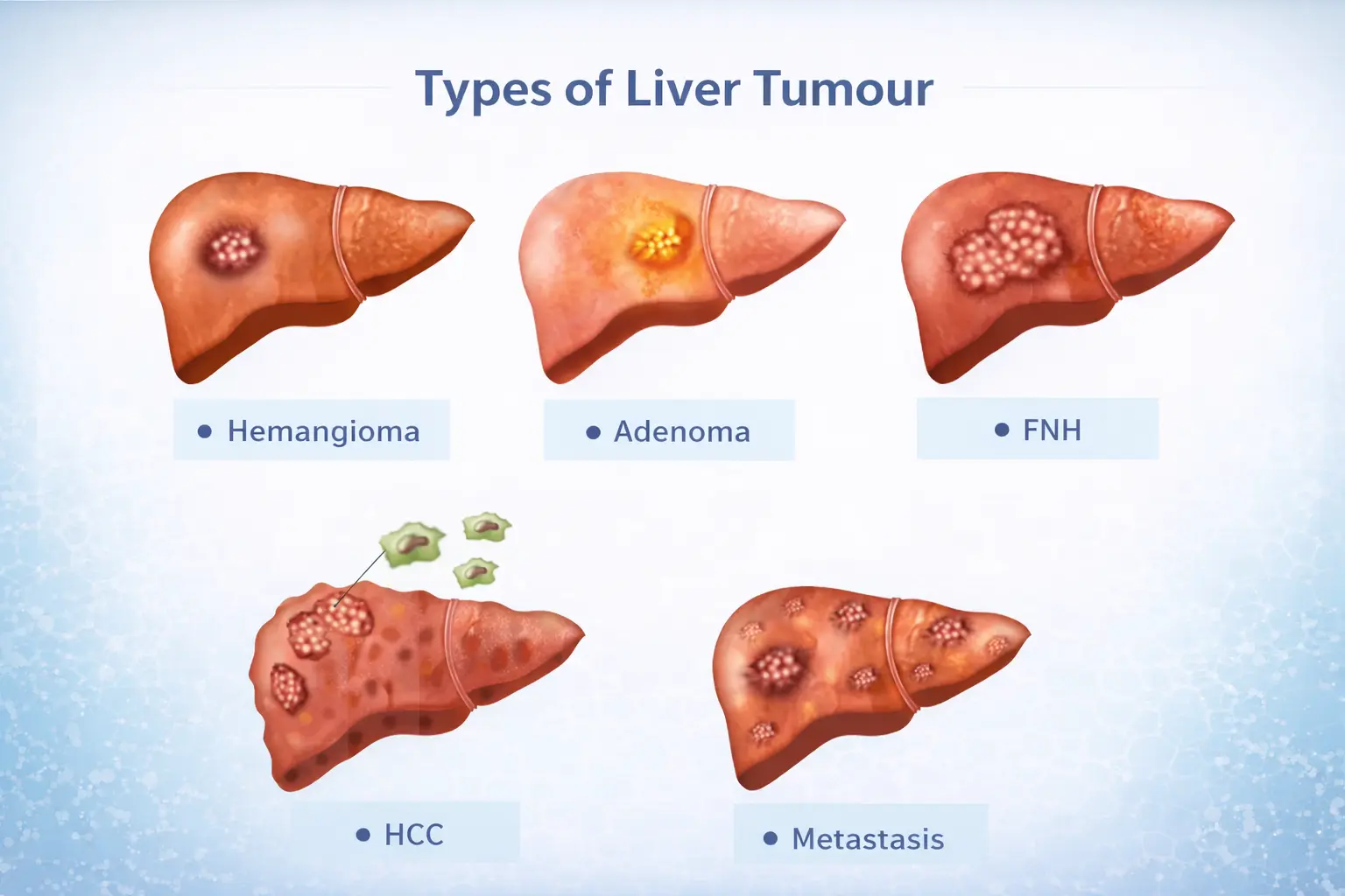
This designation refers to malignancies that originate directly within the hepatic tissue itself.
- Hepatocellular Carcinoma (HCC): Representing the predominant form, comprising approximately 75-85% of all primary hepatic cancers, this variant originates from the principal hepatic functional cells called hepatocytes.
- Intrahepatic Cholangiocarcinoma: This less common subtype arises from the epithelial tissue lining the bile conduits situated within the liver, typically exhibiting more aggressive biological behavior and yielding inferior therapeutic responses.
- Hepatoblastoma: An exceptionally uncommon variant that manifests predominantly during early childhood years.
Secondary (Metastatic) Liver Carcinoma
This form encompasses cancerous growths that have disseminated to the hepatic organ from malignancies originating in other anatomical locations, including:
- Large intestine
- Pulmonary tissue
- Mammary tissue
- Pancreatic tissue
It is noteworthy that metastatic tumors affecting the liver are considerably more prevalent than those that originate primarily within the hepatic tissue.
Staging and Disease Progression
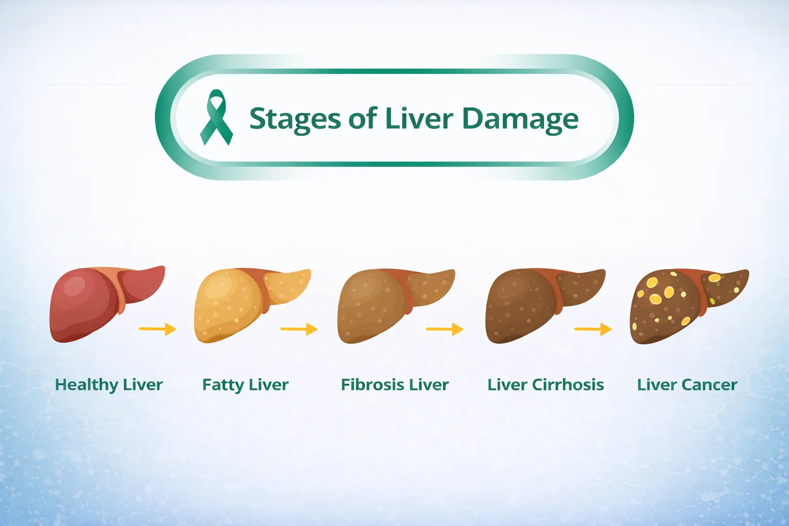
The assessment of hepatic malignancy incorporates evaluation of lesion dimensions, quantity of lesions, invasion into vascular structures, functional capacity of the hepatic tissue, and extent of dissemination throughout the body.
Early-Stage Disease
At this initial phase, lesions remain localized with modest dimensions, confined entirely to the hepatic organ. The functional integrity of the liver tissues remains substantially intact. In many circumstances, curative therapeutic approaches demonstrate favorable outcomes.
Intermediate-Stage Disease
This classification encompasses presentations with multiple lesions present, though dissemination beyond the liver has not yet transpired. However, the array of therapeutic modalities becomes more restricted in scope.
Advanced-Stage Disease
At this juncture, malignant cells have invaded the blood vessel network. Regional lymphatic tissues and distant organs have become affected by metastatic disease. Clinical manifestations become increasingly pronounced and systemic in nature.
End-Stage Disease
The hepatic organ demonstrates severely compromised functional capacity, and the malignant burden becomes extensive throughout the body. Clinical management transitions toward alleviating symptoms and maintaining quality of life through palliative approaches.
Risk Factors and Prevention
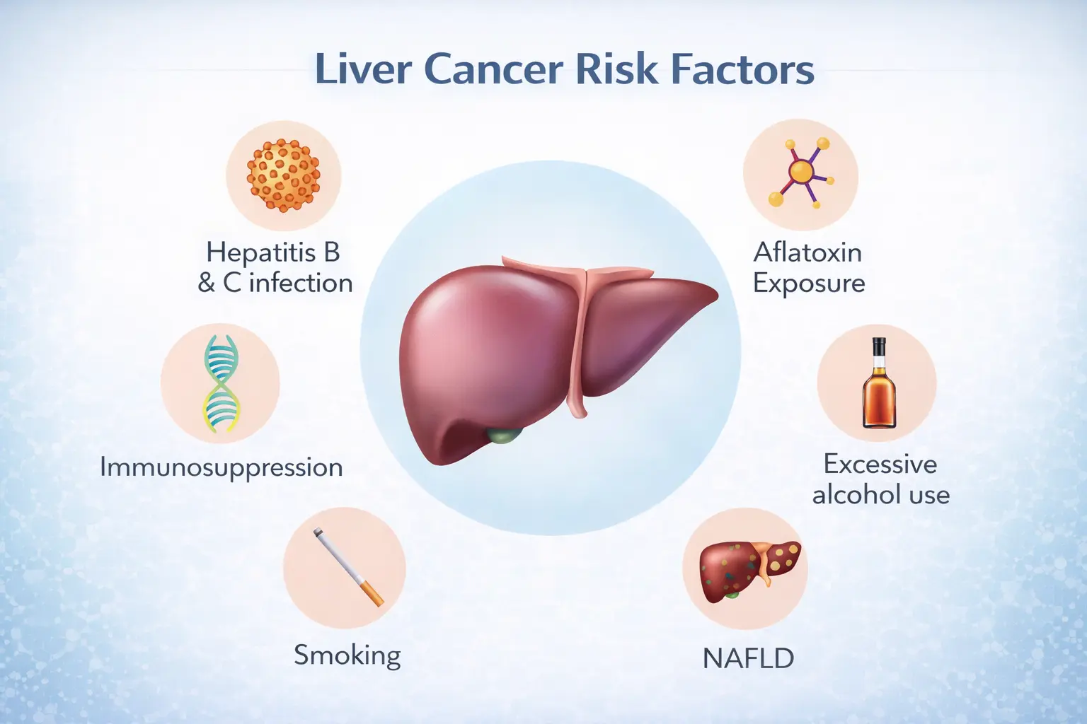
Multiple elements contribute to the development of hepatic malignancy. Individuals demonstrating one or more of these characteristics warrant heightened clinical surveillance:
- Persistent Hepatitis B Viral Infection: Long-standing infection with this pathogen represents a substantial risk determinant.
- Persistent Hepatitis C Viral Infection: Similar to HBV, chronic HCV infection significantly elevates carcinogenesis risk.
- Hepatic Fibrosis and Cirrhosis: Progressive scarring and structural degradation of hepatic tissue from any cause increases malignant transformation likelihood.
- Alcohol-Induced Hepatic Pathology: Prolonged excessive alcohol consumption causes inflammatory damage leading to cirrhotic changes and cancer development.
- Non-Alcoholic Fatty Hepatic Disorder (NAFLD): Excessive lipid accumulation within hepatocytes, often associated with metabolic syndrome, represents an emerging risk factor.
- Metabolic Conditions: Diabetes mellitus and increased body weight correlate with heightened malignancy risk.
- Mycotoxin Exposure: Consumption of contaminated foodstuffs harboring aflatoxin-producing organisms increases cancerous transformation risk.
- Tobacco Use: Smoking maintains systemic inflammatory conditions and impairs hepatic detoxification mechanisms.
- Genetic Predisposition: Family histories of hepatic malignancies suggest inherited susceptibility factors.
Clinical Manifestations
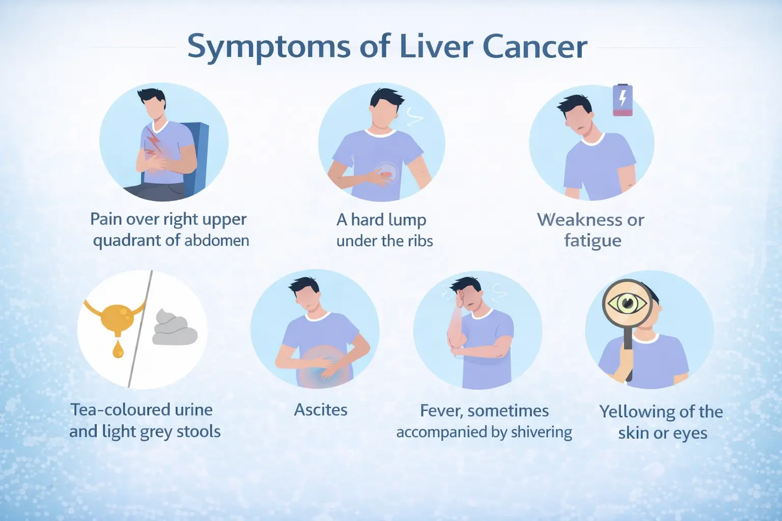
Early presentation of hepatic malignancy frequently remains asymptomatic, making early detection challenging. As pathological progression advances, affected individuals may experience:
- Discomfort or aching sensation in the right upper abdominal region
- Progressive decline in body mass without deliberate dietary restriction or increased physical activity
- Diminished appetite and reduced food intake
- Persistent exhaustion and reduced physical capacity
- Abdominal expansion resulting from accumulation of fluid (ascitic fluid)
- Yellowing of integumentary and scleral tissues (icterus)
- Sensations of gastric disturbance and emesis
- Propensity for subcutaneous hematomas and hemorrhagic manifestations from minor trauma
Diagnostic Procedures
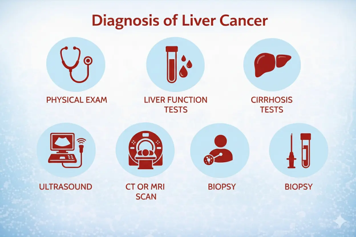
Confirmation of hepatic malignancy typically necessitates integration of multiple investigative methodologies:
Laboratory Analysis
Serological assessments evaluate specific tumor-associated proteins, particularly alpha-fetoprotein (AFP), which frequently demonstrates elevation in HCC. Comprehensive metabolic panels assess hepatic synthetic function. Serological testing determines exposure to viral hepatitis pathogens and quantifies viral replication burden in infected patients.
Radiographic Evaluation
Sonographic imaging represents an initial screening tool demonstrating excellent accessibility and reproducibility. Cross-sectional imaging via computed tomography provides superior spatial resolution for lesion characterization. Magnetic resonance imaging demonstrates exceptional sensitivity for identification of hepatic lesions and better delineates tissue characteristics.
Tissue Confirmation
Percutaneous needle aspiration or core tissue sampling confirms diagnosis when radiographic features prove ambiguous. This procedure is sometimes circumvented when imaging characteristics are distinctly pathognomonic for malignancy.
Frequently Asked Questions
Can hepatic malignancy be effectively prevented?
Yes, multiple preventive strategies substantially decrease risk. Immunological protection against Hepatitis B through vaccination provides long-term protection. Prompt therapeutic intervention for Hepatitis C infection prevents progression to cirrhosis. Moderation of alcoholic beverage consumption protects hepatic tissue integrity. Attainment and maintenance of appropriate body composition significantly reduces disease likelihood.
Does early-stage hepatic cancer respond to curative interventions?
Hepatic malignancies identified during early stages demonstrate favorable potential for curative intervention through operative resection or organ transplantation. Regrettably, advanced-stage presentations typically prove refractory to curative approaches, though various therapeutic modalities may extend survival and improve symptom burden.
Which patient populations warrant systematic screening protocols?
Individuals demonstrating persistent viral hepatitis, progressive cirrhotic changes, or recognized chronic hepatic diseases constitute appropriate candidates for regular surveillance imaging and laboratory monitoring to facilitate early diagnosis.
What determines the velocity of malignant cell proliferation?
Biological behavior varies considerably across individual tumors. Certain lesions demonstrate indolent growth trajectories with minimal clinical progression. Conversely, other malignancies display rapid expansion and tissue invasion with aggressive clinical courses.
Does hepatic malignancy typically produce somatic discomfort?
Physical discomfort typically emerges only during progressive disease stages when lesion volume increases or metastatic spread occurs. Early-stage malignancies often remain completely asymptomatic despite active cell proliferation.
Traditional Treatment Approaches
Operative Management
Surgical procedures encompass complete extirpation of malignant tissue combined with systematic lymphadenectomy of regional nodes demonstrating potential spread. This approach proves most efficacious during early disease stages when complete extirpation remains feasible.
Radiotherapeutic Intervention
High-energy particle or electromagnetic beam therapy targets hepatic lesions, inducing cellular damage incompatible with continued viability. This modality proves particularly useful for certain histological variants and advanced presentations unsuitable for operative management.
Pharmacological Chemotherapy
Systemic administration of cytotoxic pharmaceuticals represents a palliative approach for medically inoperable patients or those with distant metastatic disease. While survival extension remains modest, symptom amelioration and quality-of-life preservation constitute important clinical benefits.
Oriental Medicinal Practices
Herbal and botanical preparations from traditional Chinese medical systems demonstrate potential capacity to retard malignant progression and enhance immunological function in hepatic cancer patients. Concurrent utilization with conventional therapies may reduce adverse effects from radiotherapy and chemotherapy while simultaneously optimizing patient wellness and physiological function.
Minimally Invasive Treatment Technologies
Interventional Radiological Therapy
This technologically advanced therapeutic methodology employs real-time medical imaging guidance to accomplish percutaneous tumor treatment through minute incisions measuring merely 1-2 millimeters in width. Through image-guided needle placement, therapeutic agents or energy modalities reach the malignant tissue with exceptional precision. This approach demonstrates particular utility in hepatic malignancy management, alongside other solid tumor types including pulmonary, mammary, colorectal, cervical, nasopharyngeal, thyroid, prostate, and gastric cancers.
Cryoablative Therapy
Cryosurgical technology, alternatively termed cryoablation, represents both an ancient and modern therapeutic modality. By applying extreme cold temperatures via specially designed applicators, malignant cells undergo destruction through cytoplasmic crystallization and cellular membrane disintegration. Applicability extends across multiple solid tumors including hepatic, pulmonary, mammary, colonic, cervical, nasopharyngeal, thyroid, prostate, and gastric malignancies.
Combined Thermal Ablation System
Despite its nomenclature, this technology represents not a surgical instrument but rather an integrated cryogenic ablation platform utilizing liquid-nitrogen-based energy delivery through multiple ablation needles. The designation derives from exceptional procedural precision. This system proves beneficial for hepatic, pulmonary, pancreatic, thyroid, prostatic, renal malignancies, and various primary and secondary tumors throughout the abdomen and pelvis.
Nanoknife Irreversible Electroporation
Nanoknife technology constitutes a cutting-edge ablative methodology utilizing high-voltage electrical pulses transmitted through electrode probes. These pulses perforate tumor cell membranes with nanometer-scale precision, creating irreversible structural compromise fatal to malignant cells. This innovation proves particularly advantageous for pancreatic, hepatic, and pulmonary malignancies, especially lesions adjacent to critical anatomical structures including hilar vessels, gallbladder, bile ducts, pancreatic tissue, and urinary collecting systems.
Particle Seed Implantation
Particle knife technology, commonly designated as 125-Iodine seed implantation, employs microscopic radioactive iodine particles producing short-distance gamma radiation for continuous malignant cell elimination. This modality successfully treats numerous primary and secondary tumors and proves particularly effective for pulmonary, prostatic, mammary, hepatic, nasopharyngeal, lingual, parotid, tonsillar, cervical, and endometrial malignancies. Implanted seeds provide sustained tumoricidal radiation effects equivalent to external beam therapies.
Drug-Eluting Microsphere Chemoembolization (DEB-TACE)
This interventional procedure involves injection of microspheres composed of polymeric or ceramic materials that simultaneously deliver encapsulated anti-tumor pharmaceuticals while occluding blood vessels supplying malignant tissue. The dual mechanism—pharmaceutical delivery combined with vascular obstruction—produces superior therapeutic efficacy. Applicability encompasses various solid malignancies including hepatic, pulmonary, mammary, colonic, cervical, nasopharyngeal, prostatic, and gastric cancers.
Radiofrequency Ablation
Radiofrequency ablation (RFA), a minimally invasive therapeutic modality, employs image-guided thermal energy delivery to achieve selective tumor destruction. Under real-time radiographic visualization, energy application generates intense localized heat incompatible with malignant cell survival while sparing surrounding normal tissue. This technique proves particularly valuable for hepatic, pulmonary, mammary malignancies and various secondary metastatic tumors.