


Radiofrequency Ablation (RFA) for Cancer Treatment
What Is Radiofrequency Ablation?
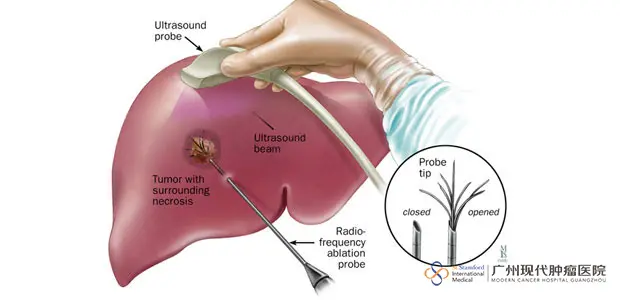
Radiofrequency ablation, commonly abbreviated as RFA, is a minimally invasive cancer treatment that relies on medical imaging to guide the destruction of tumor cells through heat. Rather than requiring an open surgical incision, the procedure uses a fine needle inserted through the skin to deliver a heat-generating probe directly into the tumor. Because the energy is concentrated precisely at the tumor site, the surrounding healthy tissue is largely spared from damage. Since RFA can achieve outcomes comparable to those of conventional surgical removal without the need for an operating incision, it is sometimes described as a “scalpel without surgery.”
How Does RFA Work?
The RFA system combines a specialized needle electrode with a multipoint temperature-monitoring mechanism, and the device is capable of generating temperatures as high as 125°C at the treatment site. Throughout the procedure, the targeted area is continuously monitored in real time to help ensure the treatment stays safe and controlled. Physicians perform the procedure with the assistance of imaging technology such as digital subtraction angiography (DSA), CT scans, or type-B ultrasound.
The needle electrode used in RFA has ten small prongs at its tip. Once the electrode is guided through the skin and into the tumor, these prongs deploy outward to anchor firmly within the tumor tissue. A high-frequency electrical current is then passed through the prongs, causing the surrounding ions to vibrate rapidly and generate friction-based heat. As the temperature within the tumor rises to somewhere between 80°C and 100°C, the heat induces coagulation necrosis, effectively destroying a portion of the cancerous cells.
What Does the RFA Procedure Involve?
Because RFA is an image-guided, minimally invasive procedure, it is typically carried out by an interventional radiologist who has received specialized training, usually within an interventional radiology suite, though it can occasionally take place in a standard operating room.
Before the Procedure
- The patient lies down on the procedure table and may be connected to monitoring equipment that tracks heart rate, blood pressure, and pulse throughout the session.
- A nurse or technologist places an intravenous line in the patient’s hand or arm to allow sedative medication to be administered as needed.
- The skin area where the electrode will be inserted is cleaned and sterilized, then covered with a surgical drape.
- If the patient will remain awake during treatment, the physician numbs the insertion site using a local anesthetic.
During the Procedure
- Guided by real-time imaging, the physician threads the needle electrode through the skin and advances it until it reaches the tumor.
- Once positioned correctly, radiofrequency energy is delivered through the electrode. Larger tumors may require the electrode to be repositioned several times so that multiple areas of the tumor can be treated, reducing the risk of leaving untreated tissue behind.
- After treatment is complete, the electrode is withdrawn, gentle pressure is applied to control any bleeding, and the puncture site is covered with a dressing. No stitches are required.
After the Procedure
The intravenous line is removed once the treatment concludes. A single ablation session generally takes between 10 and 30 minutes, though additional time is needed when several ablations are performed in one visit. In total, most RFA sessions are completed within one to three hours.
How Should Patients Prepare for RFA?
Patients should give their physician a full account of any medications they are currently taking, including herbal or over-the-counter supplements, as well as any known allergies — particularly to local anesthetics, general anesthesia, or iodine-based contrast agents (sometimes called “x-ray dye”). Depending on the individual case, the physician may recommend temporarily discontinuing aspirin, nonsteroidal anti-inflammatory drugs (NSAIDs), or blood-thinning medications ahead of the procedure.
Blood tests are typically performed beforehand to check kidney function and confirm that clotting is within a normal range.
Women should always let their physician and radiology technologist know if there is any chance they could be pregnant, since many imaging procedures are avoided during pregnancy to limit the fetus’s exposure to radiation. When an x-ray cannot be avoided, extra precautions are taken to minimize radiation exposure to the baby.
Patients are usually asked to avoid eating or drinking anything after midnight before their scheduled procedure, though their doctor will clarify which medications, if any, can still be taken that morning. It is also recommended that patients arrange for a friend or family member to drive them home afterward, and a hospital gown may be provided for the procedure itself.
What Are the Clinical Applications of Radiofrequency Ablation?
RFA is widely used in the management of liver cancer, most notably for two forms of the disease: hepatocellular carcinoma, which originates directly in the liver, and metastatic liver tumors that have spread from an original colon cancer.
The treatment tends to be most effective against primary liver tumors that measure less than 5 cm in diameter, with no more than three tumors present, or tumors smaller than roughly one and a half inches across.
RFA may be used alongside chemotherapy or radiation therapy, or as an alternative to surgery altogether. It is often considered a strong treatment option in situations such as:
- The tumor is located somewhere difficult to reach surgically, making the patient a poor candidate for an operation.
- Other existing health conditions make surgery an especially risky choice.
- Removing the tumor surgically would leave too little healthy liver tissue for the organ to continue functioning properly.
- Liver tumors have failed to respond to chemotherapy, or have returned after a previous surgical removal.
- Multiple small liver tumors are scattered in a pattern that makes surgical removal impractical.